by Dr. Nicholas Studholme, DC, CCSP, CCEP, FAFS
To say one area of the spine is more important than another would be unfair to the rest of the spine; however, it is clear that when we closely inspect the thoracic spine, it is profoundly different than the cervical or lumbar spine. It typically has twelve segments, many more than the other spinal regions, and it has a ribcage attached to it, providing significant stability and support. It also is located between the cervical and lumbar regions so any bottom‐up or top‐down movements will be forced to go through the thoracic spine.
One of the most important principles of Applied Functional Science (AFS) is gravity, and in our daily lives, the thoracic spine is constantly fighting this tremendous force. Generally, all daily movements require that we have our hands pronated, thereby constantly shortening our pecs and lats, and also create a stretch and inhibition of our scapular stabilizers (traps, rhomboids, serrratus anterior, etc.). As a result, we tend to hunch forward and yet because we have to see the horizon, we look up, thus creating anterior head carriage. This can result in significant sub‐occipital and cerico‐thoracic pain as these areas are now taking on excessive load to compensate for the rounded thoracic spine.
If we understand the mechanics of the thoracic spine, then we can use the principles of AFS to assist our patients in creating meaningful, sustainable changes. First, we must understand coupled motion, which requires nothing more than the knowledge that any movement of the spine in one plane is normally accompanied by a compatible spinal movement in another plane. A common example used is that spinal lateral flexion is always accompanied by spinal rotation. In other words, two types of motions are "coupled" together. Type 2 Motion is defined by the joints rotating and laterally flexing the same direction; Type 1 Motion is defined by the joints rotating and laterally flexing in opposite directions). The thoracic spine tends to exhibit Type 2 Motion from T1‐T5 and Type 1 from T6‐T‐12. It is theorized that when a spinal section (or an individual vertebral segment) moves in two directions that are not the expected coupled movements, then this is considered to be uncoupled mechanics. Uncoupled mechanics in spinal sections or in a vertebral segment can lead to abnormal ranges of motion, recurrent joint dysfunction, joint degeneration, inflammation, and pain.
However, when we look at many athletic endeavors, we realize that both coupled and uncoupled motions occur all the time. Therefore, we need to assess, mobilize, and train our patients and clients to be successful in all motions to avoid injury and enhance performance. When treating the thoracic spine, I always use the AFS principle of starting with success and building on success. For a majority of thoracic spine conditions, success is typically that our patients have great movement into flexion and dysfunctional extension. If we understand that all movements are three‐dimensional and understand the concept of relative joint motion, then we can create a strategy that drives motion that encourages flexion with side bending and rotation, and as we return from flexion to our starting position, we remarkably are creating thoracic joint extension. Again, keeping the patient in a successful movement pattern allows for chasing the endgame of better extension.
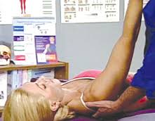
A great case example is the nursing mother patient who presents significant neck and upper thoracic and rib pain, who has to constantly hold her newborn, and who additionally has an increase in breast tissue due to nursing. This patient is permanently in an anterior head carriage neck position, has rounded shoulders, and has a more anterior center of mass. What this patient does not know is that her pain is rarely due to the neck and more often due to the thoracic spine. A typical progression in my office is to manually work tissue, then mobilize through adjustment(s) and Functional Manual Reaction (FMR), and to stabilize with matrices (three‐dimensional, logical movement patterns). For this example, I would use manual adjustments, combined with FMR in Type 1 Motion and Type 2 Motion of the thoracic spine with the pelvis in and out of synch with relationship to the shoulders (in the TrueStretch™). This would then be followed by the patient performing anterior lunges (beginning with both arms extended in front of his/her body at shoulder height) and reaching both hands in front of the lunging knee (or even in front of the lunging foot at ground height). This drives flexion of the thoracic spine as the patient lunges and creates extension of the thoracic spine as the patient returns from the lunge. If this is successful, we then go to three‐dimensional waist to shoulder dumbbell press, and then to a three‐dimensional shoulder to overhead press. Finally, if we are having success, we will ultimately finish with a Thoracic Spine Matrix.
Please review FMR of the Thoracic Spine (Functional Video Digest Series v3.10) and Thoracic Spine (Functional Video Digest Series v1.8) for more specifics pertaining to Dr. Studholme’s explanation of treatment.


